
On 18 June 2026 Midjourney, until now known for image generation, announced a whole-body ultrasound scanner that hands reconstruction and segmentation to learned models (Midjourney Medical, official post on midjourney.com/medical). It is worth reading with a technical eye, because the announcement puts together three things of different natures: a verifiable hardware choice, a plausible compute pipeline, and a set of launch claims that at this point have no independent data behind them.
Context and stated architecture
The device is a tank of warm water in which a person descends slowly, about 5 cm per second, along a ring of piezoelectric elements that act as both emitter and receiver. The aim is to scan the whole body in around sixty seconds. On the hardware side the most concrete specification is the source: the sensing comes from Butterfly Network’s ultrasound-on-chip technology, integrated as silicon modules. Butterfly confirmed its role in a separate release, citing a co-development agreement worth around 74 million dollars over five years (radiologybusiness.com).
This is the most technically relevant detail, because it sets the system’s real physical limits. Butterfly’s CMUT (Capacitive Micromachined Ultrasonic Transducer) was designed for portable point-of-care ultrasound, not for tomography. Midjourney is therefore not promising a new sensor: it is promising a different way of using many of them, arranged in a circle around a body immersed in water. The water acts as a coupling medium of known acoustic impedance, and the ring geometry gives insonification from many angles: this is the principle of ultrasound tomography, studied for decades, mostly for the breast (USCT, Ultrasound Computed Tomography).
The reconstruction pipeline
The point where physics becomes an image is the compute. A wave crossing water and tissue changes amplitude, phase and time of flight at every change in density and stiffness. By recording the returns from many angles you invert the problem and reconstruct a volumetric map of the acoustic properties. It is an expensive, ill-conditioned inverse problem: the classical methods (full-waveform inversion) iterate over high-dimensional models, and this is where learned models come in, both as reconstruction priors and to speed up the inversion.
On top of reconstruction sits a second model, for segmentation, which identifies internal structures automatically. The important distinction, and Midjourney puts it in writing, is that the initial output is body-composition maps, not diagnostic reports. Technically the distinction is sharp. Separating fat, muscle and organ outlines is a task where image segmentation models are by now mature; giving a clinical judgement about a lesion is a different class of problem, with entirely different sensitivity, specificity and validation requirements.
The critical point: the physical limits of ultrasound
The constraint no software pipeline gets around is the interaction between acoustic waves and air or bone. The acoustic impedance of air is lower than that of soft tissue by more than three orders of magnitude: at a tissue-air interface almost all the energy is reflected. That is why ultrasound does not see into the aerated lung and struggles with bowel gas. Bone absorbs and scatters heavily, and shadows whatever lies behind it. A ring geometry in water softens part of the problem on the extremities and on superficial tissue, but does not remove it for the chest, the deep abdomen and retroperitoneal structures.
So whole-body image quality comparable to magnetic resonance imaging, in a fraction of the time, should be taken for what it is: a launch statement, not a measured result. MRI has no air-and-bone problem; the comparison holds at most over specific regions, and it needs to be demonstrated with published metrics and ground truth, not with concept renders. The trade press that covered the announcement was explicit: the claims of quality superior to MRI are company framings not independently verified (radiologybusiness.com, iatrox.com).
Systemic implications
Regardless of how well any single specification holds, the technical signal is that the marginal cost of acquiring images is falling. When the transducer is a chip out of a foundry and reconstruction runs on general-purpose clusters, imaging becomes a problem of software, data and compute more than of dedicated apparatus. The practical consequence is that the bottleneck shifts: producing the image stops being the hard part.
The hard part becomes everything around it. Acquiring a whole-body volume inherently produces many incidental findings in asymptomatic people, the large majority benign, and each can trigger anxiety, work-up and procedures that carry their own risk. Whole-body screening in a healthy population has no solid evidence of benefit on mortality and outcomes today, and guidelines do not recommend it broadly. Producing large volumes of scans without a downstream path for interpretation, follow-up and clinical responsibility shifts the load rather than reducing it. This is a system problem, not a sensor one.
There is also a regulatory-positioning question. Launching as a wellness product that returns body-composition maps avoids facing the full medical-device pathway at once, with a stated intention to submit results to the FDA over time to unlock diagnostic capabilities. The boundary between a composition map and information the user perceives as clinical is thin, and it decides who carries responsibility for what a person does after seeing their own scan.
Limits of this analysis
The above rests on the launch post and the trade coverage as of 18 June 2026; there are as yet no peer-reviewed publications, reconstruction metrics, diagnostic-accuracy studies or prototype validation documentation. The roadmap figures — the first spa in San Francisco around late 2027, a fleet of over 50,000 scanners by 2031, up to a billion scans a month — and the statements about potential health benefit are announced targets, not results. Between a prototype that produces a plausible image and a medical device whose accuracy is demonstrated on a defined population lie clinical studies, independent validation and care pathways: it is that distance, not the physics of the sensor, that decides whether a technology like this reaches practice.
- https://www.midjourney.com/medical
- https://radiologybusiness.com/topics/healthcare-management/healthcare-economics/ai-lab-midjourney-investing-over-74m-launch-whole-body-ultrasound-screening-business
- https://www.iatrox.com/blog/midjourney-medical-full-body-ultrasound-scanner-explained
- https://www.noze.it/en/insights/midjourney-medical/
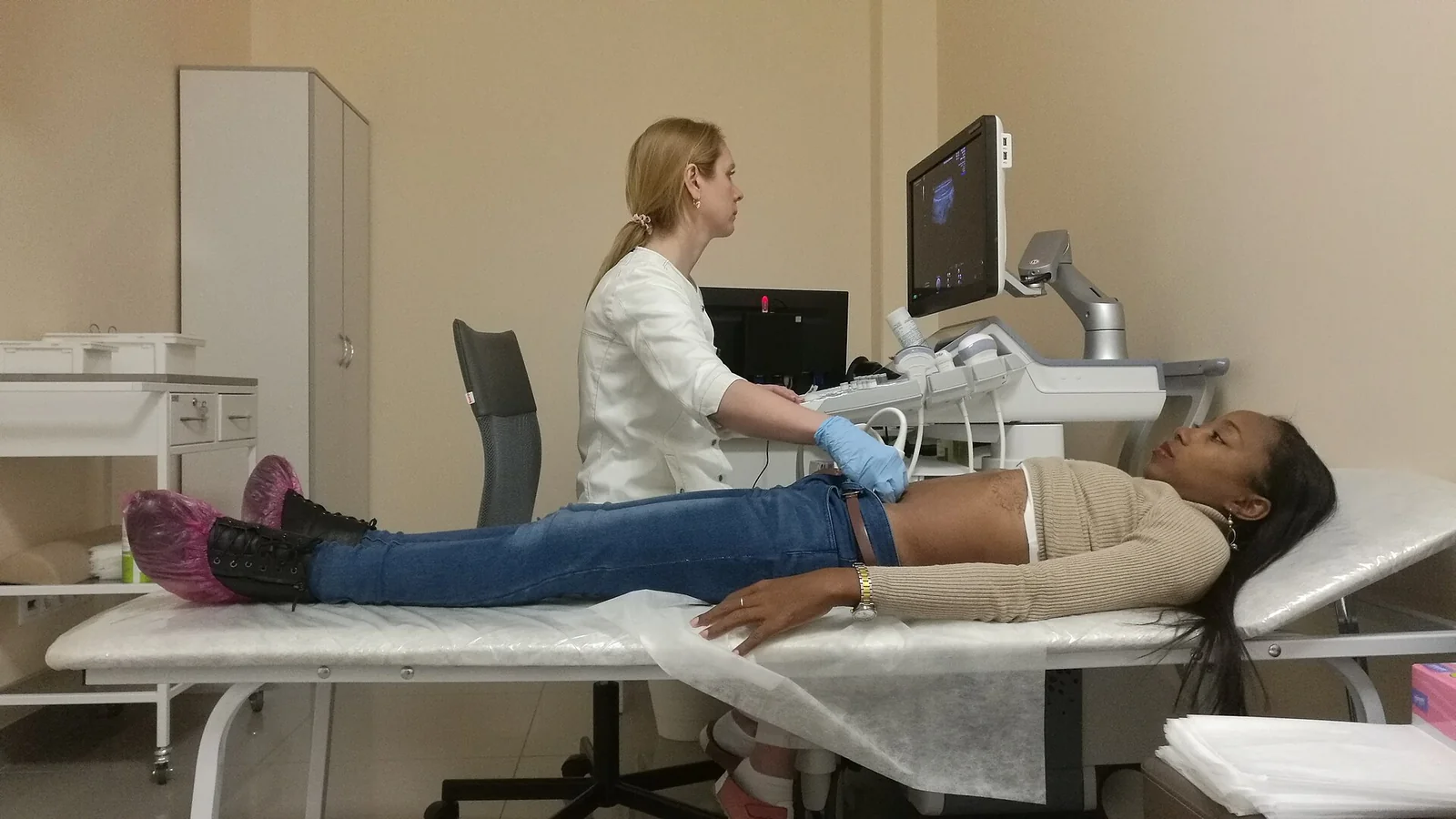
Cover image: A gloved clinician performs an abdominal ultrasound on a young woman lying on an examination couch, with the ultrasound machine and a… — photo by RG72, CC BY-SA 4.0 — https://commons.wikimedia.org/wiki/File:Kuracistino_ekzamenas_per_ultrasono_abdomenon_de_juna_virino_ku%C5%9Danta_sur_benko_en_kliniko_(Tjumeno).jpg